Detached retina recovery and outlook
Retinal detachment surgery is often very successful. Many people go back to having a similar level of sight to what they had before. But, like any surgery, it comes with risks ‑ and your vision may not be quite as good as it was.
The effectiveness of detached retina surgery depends on how much of the retina had pulled away, for how long, and if the macula (the central part of your retina) was affected. Your post‑surgery recovery can also have a big impact ‑ especially if you need to do posturing (see below). Sometimes, people need to have detached retina surgery a few times to fully treat the problem.
Vitrectomy
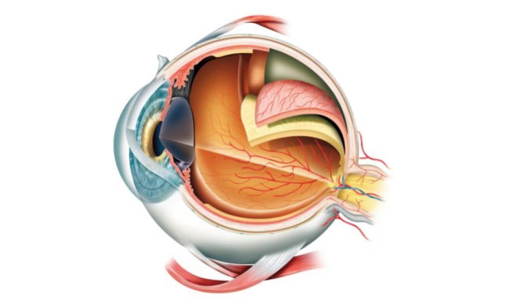
This is the most commonly used treatment for retinal detachment in Australia. The surgeon will extract some of the fluid from inside your eye and replace it with a small bubble of gas (or sometimes a type of oil). The gas bubble presses gently against the retina, helping it move back into place against the inside wall of the eye so it can reattach.
After a few weeks the gas will be absorbed by your body, and you will produce new vitreous fluid to replace what the surgeon removed.
After surgery, you may need to follow a detached retina recovery routine called 'posturing' for a couple of weeks. This involves keeping your head in specific positions ‑ like lying on your side, face down or sitting upright ‑ so the gas bubble stays in the right spot. This can be quite uncomfortable and inconvenient. But remember that it’s temporary and plays a key role in helping your eye heal properly.
If you’ve had a vitrectomy, you must avoid changes in air pressure, since this can cause the gas bubble to expand or shift, which can be dangerous. This means you should not fly or do things like scuba diving until your doctor gives you the go‑ahead.
Pneumatic retinopexy
This is a less common kind of retinal detachment surgery. As with a vitrectomy, a small amount of gas is put into your eye. But no vitreous fluid gets removed The bubble then presses the retina back in place, and cryotherapy or laser is applied around the hole or tear. The gas is reabsorbed over time and is replaced by fluid as the eye heals.
Again, if you've had gas put in your eyes, you must not fly or scuba dive until your doctor says it's OK to do so.
Scleral buckle
The sclera is the white outer layer of your eye. In scleral buckle surgery, a tiny band (often made of silicone) is placed around your eyeball. This squeezes it slightly, so the inside of your eye moves inwards. That in turn helps the retina reattach. The buckle will be left in place permanently, but it won't be visible.
Cryotherapy or laser surgery
If your retinal detachment is caused by a hole or tear in the retina, the surgeon might use a laser or cryotherapy (a freezing technique) to seal up the edges of the tear or hole. This prevents more vitreous fluid from seeping out.
'%3e%3cpath%20d='M706.453%20375.999C860.234%20375.999%20984.899%20291.829%20984.899%20188C984.899%2084.1703%20860.234%200%20706.453%200C552.671%200%20428.006%2084.1703%20428.006%20188C428.006%20291.829%20552.671%20375.999%20706.453%20375.999Z'%20fill='%23009B4F'/%3e%3cpath%20d='M278.447%20375.999C432.229%20375.999%20556.893%20291.829%20556.893%20188C556.893%2084.1703%20432.229%200%20278.447%200C124.665%200%200%2084.1703%200%20188C0%20291.829%20124.665%20375.999%20278.447%20375.999Z'%20fill='%23009B4F'/%3e%3cpath%20d='M141.484%20166.68C125.01%20162.158%20114.996%20158.927%20114.996%20150.529C114.996%20143.422%20122.102%20138.9%20133.085%20138.9C144.068%20138.9%20155.051%20143.099%20164.095%20150.529L164.418%20150.852L165.388%20129.855C156.666%20125.01%20147.944%20121.78%20133.731%20121.78C123.394%20121.78%20114.027%20124.364%20107.243%20129.209C99.8137%20134.378%2095.9374%20142.13%2095.9374%20151.175C95.9374%20168.941%20110.796%20176.694%20123.394%20179.924C140.192%20184.446%20151.821%20188.323%20151.821%20198.659C151.821%20207.381%20144.068%20212.872%20131.47%20212.872C118.872%20212.872%20106.274%20207.381%2096.5834%20198.013L96.2604%20197.69L94.3223%20219.979C104.336%20226.762%20116.934%20230.316%20130.501%20230.316C142.13%20230.316%20152.144%20227.408%20159.25%20221.917C167.003%20216.103%20171.202%20207.704%20171.202%20197.367C171.202%20181.862%20161.511%20171.848%20141.484%20166.68Z'%20fill='white'/%3e%3cpath%20d='M264.557%20188C264.557%20215.78%20246.467%20230.316%20225.471%20230.316C215.457%20230.316%20207.381%20226.762%20201.244%20221.271V253.896H182.186V146.976H201.244V156.02C207.704%20149.883%20216.103%20145.684%20226.763%20145.684C247.436%20145.36%20264.557%20160.22%20264.557%20188ZM245.498%20188C245.498%20172.494%20236.131%20162.158%20222.887%20162.158C214.811%20162.158%20207.058%20165.711%20201.567%20173.787V203.182C207.058%20210.288%20214.488%20213.841%20222.887%20213.841C236.131%20213.841%20245.498%20203.505%20245.498%20188Z'%20fill='white'/%3e%3cpath%20d='M352.095%20194.137H294.274C296.535%20207.058%20305.903%20213.841%20318.824%20213.841C329.807%20213.841%20337.882%20211.257%20346.604%20206.089L347.573%20222.886C339.82%20227.085%20330.13%20230.316%20317.532%20230.316C294.274%20230.316%20274.893%20215.78%20274.893%20188.323C274.893%20162.804%20294.274%20145.683%20317.209%20145.683C341.112%20145.683%20353.71%20160.22%20353.71%20181.216C353.71%20185.415%20353.064%20189.615%20352.095%20194.137ZM335.621%20178.632C335.621%20168.618%20329.161%20160.866%20317.532%20160.866C306.549%20160.866%20296.858%20168.295%20294.597%20180.893H335.298C335.621%20180.247%20335.621%20179.601%20335.621%20178.632Z'%20fill='white'/%3e%3cpath%20d='M443.189%20221.917L445.128%20203.505C452.88%20209.965%20463.54%20213.841%20474.846%20213.841C482.921%20213.841%20488.413%20210.611%20488.413%20204.797C488.413%20189.615%20444.482%20197.367%20444.482%20169.91C444.482%20153.436%20458.695%20145.36%20475.492%20145.36C487.444%20145.36%20496.488%20148.268%20503.272%20152.467L502.303%20169.587C494.227%20164.096%20484.213%20161.189%20475.492%20161.189C468.708%20161.189%20463.217%20163.773%20463.217%20168.941C463.217%20181.862%20507.471%20175.402%20507.471%20203.828C507.471%20221.271%20492.289%20229.993%20474.523%20229.993C461.602%20230.316%20450.942%20227.085%20443.189%20221.917Z'%20fill='white'/%3e%3cpath%20d='M590.488%20177.986V228.701H571.429V220.302C564.969%20226.439%20556.57%20229.993%20546.88%20229.993C528.467%20229.993%20517.484%20219.01%20517.484%20203.505C517.484%20187.03%20530.405%20177.017%20549.464%20177.017C556.57%20177.017%20563.677%20178.309%20571.752%20180.57V177.986C571.752%20165.388%20562.385%20159.897%20550.756%20159.897C542.034%20159.897%20532.02%20163.127%20524.914%20168.941L524.268%20152.467C530.405%20148.268%20541.711%20145.037%20554.309%20145.037C574.983%20145.36%20590.488%20155.374%20590.488%20177.986ZM571.429%20206.735V194.46C564.646%20192.199%20558.185%20190.907%20551.402%20190.907C541.711%20190.907%20535.574%20196.075%20535.574%20203.505C535.574%20210.934%20542.034%20215.78%20550.433%20215.78C558.508%20215.78%20564.969%20212.872%20571.429%20206.735Z'%20fill='white'/%3e%3cpath%20d='M592.104%20146.976H612.777L636.358%20198.659L659.616%20146.976H680.289L641.526%20228.701H630.866L592.104%20146.976Z'%20fill='white'/%3e%3cpath%20d='M754.907%20194.137H697.409C699.67%20207.058%20709.038%20213.841%20721.959%20213.841C732.941%20213.841%20741.017%20211.257%20749.739%20206.089L750.708%20222.886C742.955%20227.085%20733.264%20230.316%20720.667%20230.316C697.409%20230.316%20678.027%20215.78%20678.027%20188.323C678.027%20162.804%20697.409%20145.683%20720.343%20145.683C744.247%20145.683%20756.845%20160.22%20756.845%20181.216C756.522%20185.415%20756.199%20189.615%20754.907%20194.137ZM738.756%20178.632C738.756%20168.618%20732.295%20160.866%20720.667%20160.866C709.684%20160.866%20699.993%20168.295%20697.732%20180.893H738.433C738.433%20180.247%20738.756%20179.601%20738.756%20178.632Z'%20fill='white'/%3e%3cpath%20d='M826.618%20148.914L824.034%20166.357C819.511%20163.773%20814.989%20163.127%20810.467%20163.127C802.068%20163.127%20794.315%20167.326%20788.824%20176.371V228.701H769.766V146.976H788.824V157.958C794.315%20150.529%20802.068%20145.36%20812.728%20145.36C817.896%20145.36%20822.418%20146.653%20826.618%20148.914Z'%20fill='white'/%3e%3cpath%20d='M832.109%20221.917L834.048%20203.505C841.8%20209.965%20852.46%20213.841%20863.766%20213.841C871.841%20213.841%20877.333%20210.611%20877.333%20204.797C877.333%20189.615%20833.401%20197.367%20833.401%20169.91C833.401%20153.436%20847.615%20145.36%20864.412%20145.36C876.364%20145.36%20885.408%20148.268%20892.192%20152.467L891.223%20169.587C883.147%20164.096%20873.133%20161.189%20864.412%20161.189C857.628%20161.189%20852.137%20163.773%20852.137%20168.941C852.137%20181.862%20896.391%20175.402%20896.391%20203.828C896.391%20221.271%20881.209%20229.993%20863.443%20229.993C850.522%20230.316%20839.862%20227.085%20832.109%20221.917Z'%20fill='white'/%3e%3cpath%20d='M429.298%20206.412C422.838%20211.257%20416.054%20213.841%20408.302%20213.841C395.058%20213.841%20383.429%20204.474%20383.429%20188C383.429%20171.525%20395.058%20162.158%20408.302%20162.158C416.054%20162.158%20422.838%20164.742%20429.298%20169.264C430.268%20163.45%20431.237%20157.958%20432.852%20152.467C426.068%20148.591%20417.67%20145.683%20406.041%20145.683C382.783%20145.683%20363.725%20161.512%20363.725%20188C363.725%20214.487%20382.783%20230.316%20406.041%20230.316C417.67%20230.316%20426.068%20227.085%20432.852%20222.886C431.237%20217.395%20429.944%20211.903%20429.298%20206.412Z'%20fill='white'/%3e%3c/g%3e%3cdefs%3e%3cclipPath%20id='clip0_1478_9029'%3e%3crect%20width='984.9'%20height='375.999'%20fill='white'/%3e%3c/clipPath%3e%3c/defs%3e%3c/svg%3e)